What’s the diagnosis?
Itchy papules on the trunk and limbs

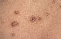
Figure 1. Multiple pale pink papules at different stages. Note the hyperpigmented areolae, increased skin markings (lichenification), and focal dark crust.

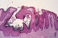
Figure 2. Epidermal hyperplasia with scant Iymphocytic inflammation and superficial dermal fibrosis
Differential Diagnosis
Papular urticaria, which are erythematous lesions with a central crust and may be caused by infestation or insect bites. The lesions are more acute, lack the accentuated skin markings and have a prominent lymphocytic infiltrate with eosinophils within the superficial and deeper dermis.
Lichen planus is characterised by itchy papules but usually these are shiny, violaceous, sharply demarcated and not associated with a central crust. Mucous membrane lesions may coexist. Skin biopsy shows a hyperplastic epidermis with a dense lymphocytic infiltrate which interacts with the basal keratinocytes at the junction.
Multiple dermatofibromas may have a firm central papule and a hyperpigmented margin but are usually nonpruritic, isolated and concentrated on the lower limbs. Skin biopsy shows a hyperplastic epidermis with underlying interlacing bundles of fibroblasts that extend into the deep dermis.
Prurigo nodules is the correct diagnosis and may develop as a distinct phenomenon or in association with atopic dermatitis, persistent insect bites or folliculitis. These papules may herald bullous pemphigoid Metabolic causes such as liver disease, renal failure, thyroid disease or lymphoma may need to be excluded. Treatments include potent topical corticosteroids or intralesional corticosteroids and antihistamines. Ultraviolet light therapy may be helpful when the lesions are widespread In severe cases oral thalidomide (available through the SAS scheme) has been helpful but is often limited by the toxicity of this drug.
A 47-year-old woman developed over a two-year period persistent itchy papules on her trunk and limbs. Each lesion had a firm pale pink central papule and a hyperpigmented margin which faded into the surrounding skin (Figure 1). Some of the papules had a central crust and the skin markings were accentuated.
Skin biopsy showed an irregularly hypertrophic epidermis with a central crater associated with scant lymphocytic inflammation and superficial dermal fibrosis (Figure 2).

