What’s the diagnosis?
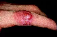
Nodular lesion with pustules on the index finger

Figure 1. Nodule with pustules on the index finger.

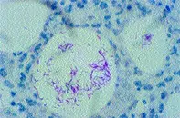
Figure 2. Skin biopsy demonstrating multiple organisms with acid-fast staining.
Differential Diagnosis
Fungal infection due to a range of dermatophytes or Sporothrix schenckii (sporotrichosis) may present as pustules and nodules rather than the more typical annular scaling lesions. Fungal scrapings may be negative, and biopsy may be required for culture or histological identification of the organisms. Fungi appear as septate hyphae or spores in the tissue, but these may be masked by intense inflammation. Oral antifungal medication is needed to treat the deep-seated fungi in this type of nodular presentation.
Resistant bacteria may also be a cause of failure of antibiotics to work. The resistance may be due to inappropriate selection of antibiotic or acquired resistance. Some cutaneous infections may be slow to respond and antibiotics may need to be taken over several months. Swabs or a biopsy of the pustular area for bacterial cultures and bacterial sensitivities may help in this situation.
Chemical or foreign body granulomas may present as persistent nodules and pustules. They may follow industrial injury or may be the result of hypersensitivity to plant or marine foreign bodies. Skin biopsy may reveal polarisable or calcified material.
Atypical mycobacterial skin infection (fish tank or swimming pool granuloma) is the correct diagnosis in this case, based on a skin biopsy that revealed multiple rod-shaped organisms in the tissue (Figure 2) and the clinical setting. Mycobacterium marinum was cultured. This infection occurs particularly in individuals who clean tropical fish tanks, but may also occur after exposure to the organisms in swimming pools, ocean beaches or lakes. The incubation period may vary from weeks to months. As well as solitary erythematous nodules with pustules, a series of nodules may develop along the lymph vessels. In immunosuppressed individuals wider dissemination may occur. Skin biopsy may fail to reveal organisms and cultures may be required to isolate the mycobacteria, or PCR analysis for tissue mycobacterial DNA can be extracted from biopsy sections.
Successful treatments have included minocycline, doxycycline, trimethoprim–sulfamethoxazole, clarithromycin, or rifampicin plus ethambutol. These antibiotics usually need to be taken for several months.
Over a five-week period, a 23-year-old man who was a tropical fish fancier developed a persistent nontender erythematous nodule with pustules on his left index finger (Figure 1). The nodule had failed to respond to a course of cephalexin.

