What’s the diagnosis?
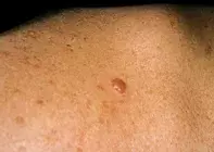
A pearly nodule on the back

Figure 1. A pearly nodule on the patient’s upper back.

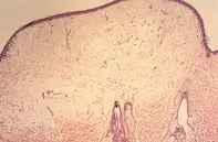
Figure 2. Skin biopsy showing elevated epidermis under which there is marked oedema with separation of collagen bundles.

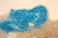
Figure 3. Colloidal iron stain demonstrating abundant mucin which stains blue and accounts for the pearly clinical appearance.
Differential Diagnosis
Basal cell carcinoma may have an identical clinical appearance and was the provisional diagnosis prior to excision. Skin biopsy of a basal cell carcinoma reveals clusters of basaloid cells forming lobules and cords in addition to the presence of mucin.
Neurofibroma presents as a flesh coloured papule but may on occasion have a more translucent quality. Skin biopsy shows numerous spindle cells associated with fibrillar stroma and variable mucin.
Cutaneous lymphoma may present as flesh coloured translucent nodules, and on skin biopsy reveals sheets of monomorphous, atypical lymphocytes in the dermis.
Focal papular mucinosis is the correct diagnosis and usually eludes clinical diagnosis. The solitary nodules typically occur on the face, trunk or proximal limbs. Stable longstanding lesions are usually not subject to biopsy, and the frequency of focal papular mucinosis is uncertain. The presence of multiple lesions should prompt a review for associated connective tissue disease, particularly lupus erythematosus or dermatomyositis as well as thyroid disease.
A 38-year-old man presented with an asymptomatic pearly nodule on his upper back (Figure 1). The nodule was sharply demarcated and had a smooth, shiny surface. The duration of the lesion was uncertain. Excision biopsy revealed an elevated epidermis under which there was marked dermal oedema with separated collagen bundles and sparse fibroblasts (Figure 2). There was no evident atypia. A colloidal iron stain showed abundant mucin (Figure 3).

