What’s the diagnosis?
A teenage girl with hyperpigmented axillary skin

Case presentation
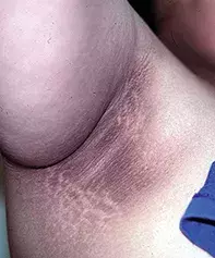
A 14-year-old girl presents with a history of skin changes, with progressive thickening and hyperpigmentation affecting both axillae (Figure) as well as her neck and groin. She is obese but appears otherwise healthy.
Diagnosis
Acanthosis nigricans is the correct diagnosis.
In the differential diagnosis, one should consider Dowling–Degos disease, a rare but harmless genetic condition that can cause hyperpigmented skin of the axillae and groin. However, the lesions tend to be macular and reticulate, without the characteristic thickening seen in acanthosis nigricans.
Pathogenesis
Acanthosis nigricans is a benign cutaneous manifestation of an underlying disorder, most commonly insulin resistance. Inappropriate activation of the insulin-like growth factor 1 receptor on the epidermis results in excess proliferation of keratinocytes and fibroblasts, which is responsible for its characteristic appearance. It presents in a high proportion of patients with endocrinopathies associated with increased insulin resistance and high insulin levels, such as obesity, type 2 diabetes and polycystic ovary syndrome. Obese individuals without an endocrinopathy may also have acanthosis nigricans, which has been termed benign acquired acanthosis nigricans. However, such patients often have high fasting insulin levels associated with obesity.
Acanthosis nigricans can also present secondary to medications that cause hyperinsulinaemia, such as prednisone, insulin and hormonal therapies. It has been reported as a complication of antiretroviral therapy.1
In rare cases, acanthosis nigricans can be associated with familial genetic mutations, either as a dominant trait or as part of a syndrome. It has also been reported in autoimmune conditions and can arise as a paraneoplastic manifestation of underlying malignancies, especially gastric cancers.2
Clinical presentations
Acanthosis nigricans is characterised by thickened, dry and rough skin that is typically grey-brown to black in appearance. The skin becomes plaque-like and velvety in texture, with or without papillomatous elevations. It commonly develops symmetrically on the axillae, neck and groin regions but can appear on any part of the skin, particularly the flexural regions. There may be associated multiple skin tags. As it becomes more severe, there is further accentuation of the skin contours and the skin becomes rugose in nature.
Most people are asymptomatic from the lesions themselves, although extremely thick lesions that become inflamed or macerated have an increased risk of secondary bacterial or fungal infection. The most severe forms of acanthosis nigricans are usually associated with underlying malignancy.
Investigations
All patients presenting with acanthosis nigricans should have a glucose tolerance test and fasting insulin levels. Referral to an endocrinologist is recommended to assess the need to investigate for other endocrinopathies. In a 14-year-old girl who is obese but otherwise healthy, investigation for an underlying malignancy would not usually be necessary.
Management
Acanthosis nigricans is a chronic condition and management should be directed at the underlying cause. In this girl’s case, achieving weight loss and implementing lifestyle changes would be most important. Treatment options for acanthosis nigricans include topical retinoids, calcipotriol, urea, salicylic acid and laser therapy, but these all tend to be disappointing and are often irritating to skin, particularly in flexural regions. Metformin has been reported to be of benefit in acanthosis nigricans secondary to insulin resistance,3,4 and this is likely to help this patient.
References
1. Mellor-Pita S, Yebra-Bango M, Alfaro-Martínez J, Suárez E. Acanthosis nigricans: a new manifestation of insulin resistance in patients receiving treatment with protease inhibitors. Clin Infect Dis 2002; 34: 716-717.
2. Berk DR, Spector EB, Bayliss SJ. Familial acanthosis nigricans due to K650T FGFR3 mutation. Arch Dermatol 2007; 143: 1153-1156.
3. Walling HW, Messingham M, Myers LM, Mason CL, Strauss JS. Improvement of acanthosis nigricans on isotretinoin and metformin. J Drugs Dermatol 2003; 2: 677-681.
4. Bellot-Rojas P, Posadas-Sanchez R, Caracas-Portilla N, et al. Comparison of metformin versus rosiglitazone in patients with acanthosis nigricans: a pilot study. J Drugs Dermatol 2006; 5: 884-889.
Dr Lee is a Dermatology Research Fellow in the Department of Dermatology, Royal North Shore Hospital, Sydney.
Associate Professor Fischer is Associate Professor of Dermatology at Sydney Medical School – Northern, University of Sydney, Royal North Shore Hospital, Sydney, NSW.
Competing interests: None.
Endocrine diseases

